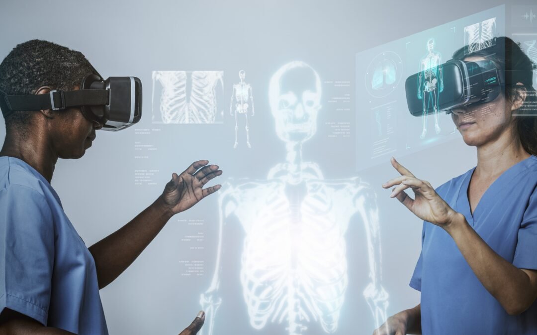
Anatomy is a three-dimensional subject that has mostly been taught in two dimensions. Students learn the course of a nerve from a diagram, memorise relations from a textbook page and get limited, precious time in the dissection hall. Virtual reality changes that balance by putting the body in front of the learner at full scale, from every angle, as often as they need.
The problem with learning a 3D body from flat pages
Ask any first-year medical student what makes anatomy hard and you will hear the same answer: spatial relationships. Knowing that the brachial plexus has roots, trunks, divisions, cords and branches is one thing. Picturing how those structures twist around the axillary artery, and what that means when a patient presents with weakness in the hand, is another.
Traditional resources each solve part of the problem. Cadaveric dissection is the gold standard for real tissue, but access is limited, specimens are expensive to maintain and a structure is only dissected once. Textbooks and atlases are portable, but flat. Plastic models are three-dimensional, but small, static and shared between many students.
What changes inside a headset
In VR, a learner can stand next to a life-size body, walk around it, scale it up until a single organ fills the room, and peel away layers one system at a time. Structures can be isolated, highlighted and labelled, then hidden again for self-testing. Because nothing is consumed, the same exploration can be repeated the night before an exam, or months later in a clinical year.
- Scale and perspective. Students see relationships from positions that are impossible with a book: from inside the thoracic cavity, or looking up through the pelvic floor.
- Layer-by-layer exploration. Skin, muscle, vessels, nerves and bone can be toggled on and off to show how systems overlap.
- Unlimited repetition. A dissection can only be done once. A virtual one can be reset in a second.
- Learning together. In a shared virtual lab, a tutor and a whole small group can stand around the same model and point to the same structure, wherever they are physically located.
VR works as a partner to the dissection hall, not a replacement. It is strongest for preparation before a practical, for revision afterwards, and for students who cannot be on campus.
Why immersion helps things stick
Educators have long observed that people remember what they do better than what they read. Immersive learning turns anatomy into something students do: they move, reach, rotate and explain. That active engagement, combined with the ability to revisit material whenever they want, is a big part of why medical schools around the world are piloting VR in their curricula.
It also changes the conversation in the room. When a tutor can say “stand here and look at where the recurrent laryngeal nerve hooks under the arch of the aorta”, the explanation and the experience happen at the same moment.
How we are approaching it at Kashmeta
Our virtual medical campus includes an anatomy lab designed for exactly this kind of guided, shared exploration. Teachers and students join as avatars, talk with spatial voice, and work around the same 3D models. Because the lab sits inside a full campus, an anatomy session can flow straight into a lecture in the auditorium or a case discussion in a meeting room, without anyone changing app.
If you lead an anatomy or medical education team and want to see what this looks like with your own curriculum, we would be happy to show you around.